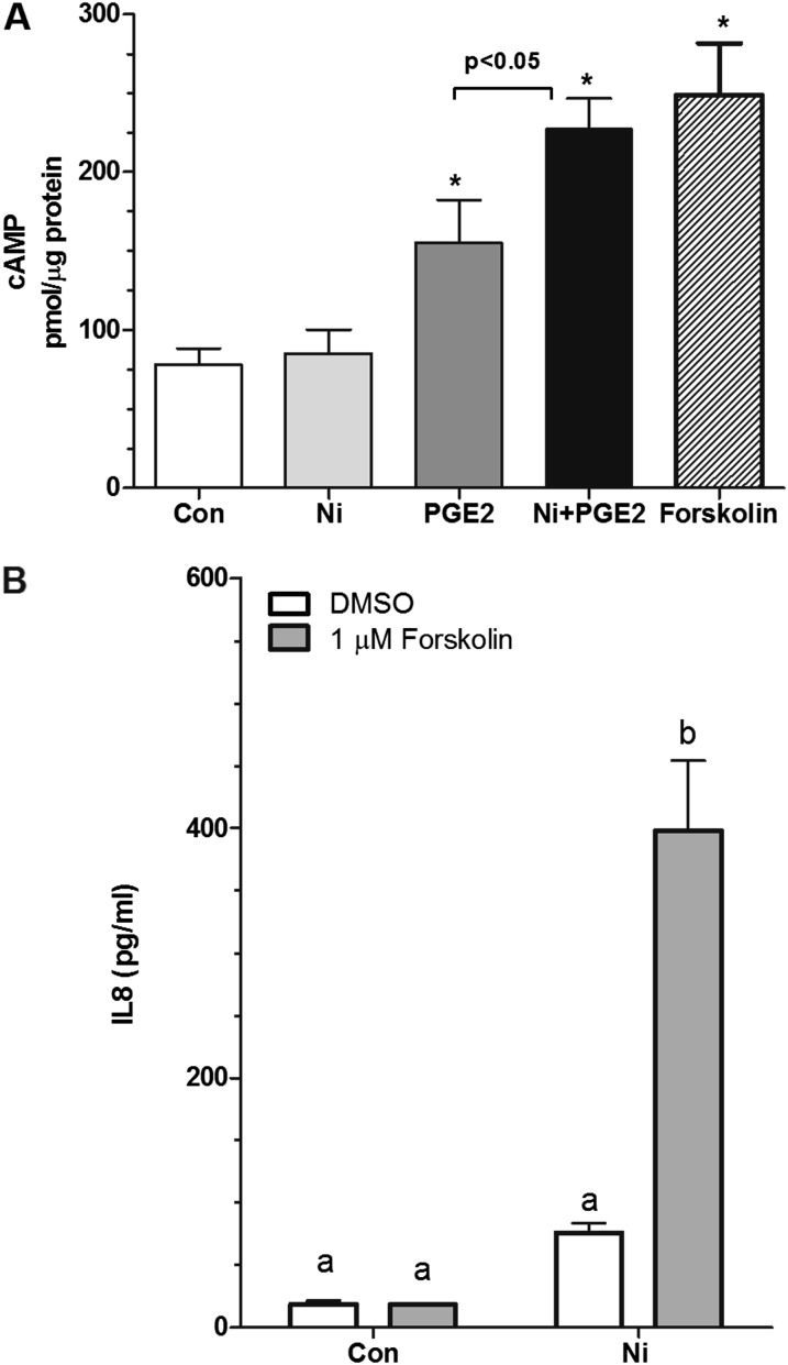
Figure 3.
PGE2 increases cAMP levels in HLFs. (A) Cells were seeded into 60-mm dishes at a density of 5.5 × 10⁴ cells/cm2 and allowed to attach overnight before stimulation with 10 nM PGE2 in the presence or absence of 200 μM Ni in serum-free modified Eagle’s medium containing 0.1% BSA for 10 minutes. Forskolin (1 μM) was used as a positive control. Activation of cAMP was determined using an enzyme immunoassay kit. Data shown are mean ± SEM pmol cAMP/μg protein (n = 4–5 dishes). *Different from control-treated cells (P < 0.05) as determined by ANOVA with Tukey’s multiple comparisons test. (B) Cells were stimulated with 200 μM Ni in the presence or absence of forskolin (1 μM) for 48 hours. Data shown are mean ± SEM and are representative of three independent experiments performed in triplicate. Points with different letters have means that are significantly different (P < 0.001) as determined by two-way ANOVA with Bonferroni multiple comparisons test.

