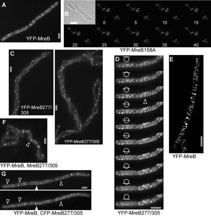
FIGURE 5:
Fluorescence microscopy of exponentially growing chains of B. subtilis cells. (A) Epifluorescence of YFP-MreB. (B) Time-lapse SIM (Zeiss; 5-s intervals) of cells expressing D158A mutant YFP-MreB; inset, outlines of cells by bright-field light. (C) Expression of double mutant YFP-MreB277/305 for 8 h, epifluorescence. (D) YFP-MreB277/305 expressed for 3 h; SIM (Zeiss) stack with planes indicated by bars in circle; triangle shows an example of an extended (<750 nm) filament. (E) SIM (Zeiss) image of cells expressing YFP-MreB under identical conditions as in D. (F) Localization of YFP-MreB 4 h after expression of double mutant MreB. Note that the helical phenotype has not fully arisen at this time, but the localization of proteins is easier to see because they are still mostly flat. White triangle indicates clear, extended filament. (G) Colocalization of CFP-MreB and YFP-MreB277/305 (examples indicated by triangles) 4 h after expression of double-mutant MreB (epifluorescence). White bars, 2 μm.

