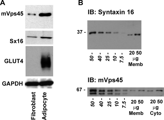
FIGURE 1:

mVps45 and syntaxin 16 levels in 3T3-L1 adipocytes. (A) Representative immunoblot of 50 μg of lysate prepared from either 3T3-L1 fibroblasts or adipocytes (as labeled) and probed with anti-mVps45, anti-Sx16, anti-GLUT4, or anti-GAPDH. Data from a representative experiment is shown, repeated three times with qualitatively similar results. Table 1 shows quantification of levels of Sx16 and mVps45 in these cells. (B) Representative immunoblot of 3T3-L1 adipocyte membranes (20- and 50-μg membranes for Sx16) or membranes and cytosol (20- and 50-μg membranes and cytosol for mVps45) compared with known amounts of recombinant Sx16 or mVps45 expressed and purified from bacteria. Figures in italics are nanograms of recombinant protein, and the position of molecular weight markers is shown. Data are from a representative experiment repeated at least four times.
