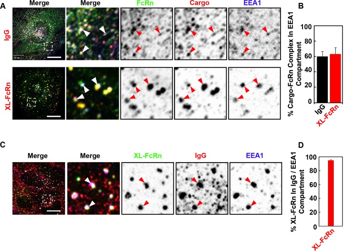
FIGURE 2:
Monomeric and cross-linked FcRn complexes are internalized into the same early endosomal compartment. (A, B) IgG or directly cross-linked FcRn is delivered to early endosomes after endocytosis. HMEC-1 cells stably expressing HA-FcRn-EGFP were incubated with either monomeric Alexa 568–human IgG (25 μg/ml) at pH 6 for 15 min at 37°C or with rat anti-HA primary antibodies (2 μg/ml) at pH 7.4 for 30 min at 37°C, followed by Alexa 568–goat anti-rat secondary antibodies (40 μg/ml) for 15 min at 37°C. (C, D) Cross-linked FcRn is internalized into early endosomes containing internalized IgG. HMEC1 cells stably expressing HA-FcRn were first incubated with rat anti-HA primary antibodies (2 μg/ml) at pH 7.4 for 30 min at 37°C, followed by coincubation with monomeric Alexa 568–human IgG (25 μg/ml) and Alexa 488–anti-rat secondary antibodies (40 μg/ml) at pH 6 for 15 min at 37°C. All samples were then fixed and stained for the early endosome antigen 1 (EEA1). Representative confocal middle sections of each condition are shown, with arrowheads indicating examples of colocalization between FcRn, ligands, and EEA1, and displayed as described in the Figure 1 legend. Scale bar, 10 μm. Bar plots show the quantification of fraction of IgG and cross-linked FcRn in the EEA1 compartment (B) or the fraction of cross-linked FcRn in IgG-containing early endosomes (D; average and SD of three independent experiments, n = 22 cells).

