FIGURE 3:
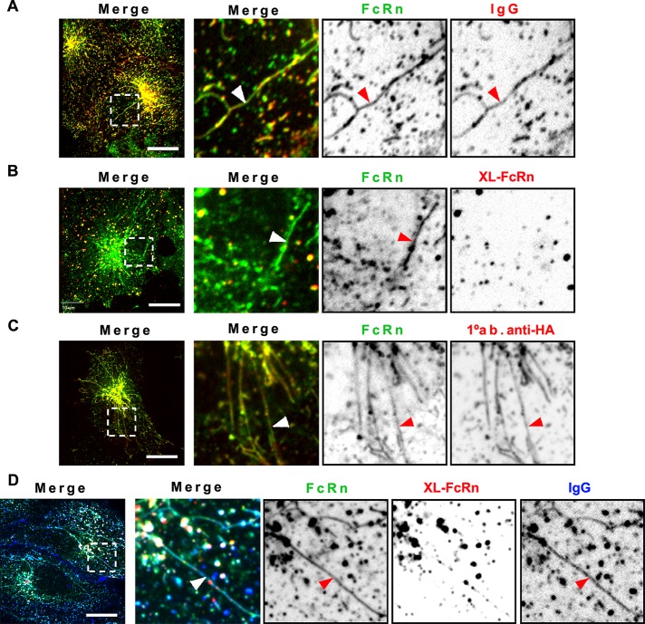
Monomeric but not multimeric IgG-FcRn complexes are sorted into recycling tubules. HMEC-1 cells stably expressing HA-FcRn-EGFP were incubated with monomeric Alexa 568–human IgG (25 μg/ml) at pH 6 (A) or Alexa 568–rat anti-HA primary antibody (25 μg/ml) at pH 7.4 for 15 min at 37°C (C). Other samples were incubated with unlabeled rat anti-HA antibody (2 μg/ml, B; 0.2 μg/ml, D) for 1 h at 37°C, followed by further incubation with Alexa 568–goat anti-rat antibody for 15 min at 37°C. Some samples were further incubated with Alexa 647–human IgG (50 μg/ml; D). All samples were then washed and transferred to an open perfusion chamber for live-cell imaging acquisition. To stimulate formation of recycling tubules, some samples (C, D) were incubated with HBSS-H (pH 7.4) containing 10 μM BFA during live-cell imaging acquisition. Time series were recorded with an interval of 2 s. Representative confocal middle sections of each condition are shown, with arrowheads indicating examples of tubules, and displayed as described in the Figure 1 legend. Note the strong increase in sorting tubules, containing IgG, or primary only antibody–FcRn complexes, and large vesicles containing cross-linked FcRn complexes, which are absent from sorting tubules. Scale bars, 10 μm.

