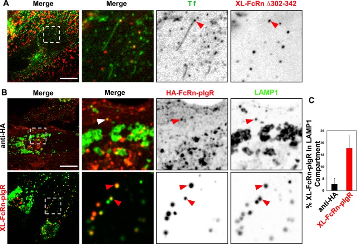
FIGURE 5:
Diversion of cross-linked FcRn to lysosomes depends on its lumenal domain. (A) Cross-linked FcRn tailless mutant is not efficiently sorted into recycling tubules. HMEC-1 cells stably expressing HA-FcRnD303-342 were incubated with rat anti-HA antibody (2 μg/ml) for 1 h at 37°C. Samples were then washed and further incubated with Alexa 488–human Tf and Alexa 568–goat anti-rat antibody for 15 min at 37°C. Samples were then washed and transferred to an open perfusion chamber for live-cell imaging acquisition, and medium containing 10 μM BFA was added. Time series were recorded immediately after the addition of BFA. Representative confocal middle sections of each condition are shown, with arrowheads indicating examples of tubules containing transferrin and lacking cross-linked HA-FcRnD303-342 multimeric complexes, and displayed as described in the Figure 1 legend. (B, C) Cross-linked HA-FcRn-pIgR chimeric receptor complexes are delivered into lysosomes. Cells stably expressing HA-FcRn-pIgR were first incubated with rat anti-HA primary antibodies (2 μg/ml) at pH 7.4 for 30 min at 37°C, followed by incubation with Alexa 568–anti-rat secondary antibodies (40 μg/ml) for 30 min at 37°C. All samples were then fixed and stained for LAMP-1. Representative confocal middle sections of each condition are shown, with arrowheads indicating examples of colocalization between FcRn-pIgR and LAMP-1, and displayed as described in the Figure 1 legend. Scale bars, 10 μm. (B) Quantification of fraction of cross-linked FcRn-pIgR in LAMP-1 (average and SD of three independent experiments, n = 27 cells).

