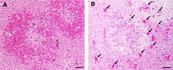
Figure 6.

Immunohistochemical staining for cleaved caspase-3 in the area of haemorrhage in the brains of fatal CM cases. (A) Representative photograph of area of congestion and ring haemorrhage is shown with H&E stain. (B) Many glial cells located around and within the area of haemorrhage show strong positive staining for cleaved caspase-3 (arrows). Scale bars = 50 μm.
