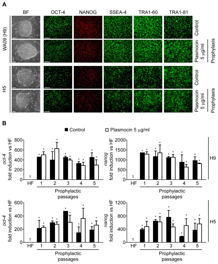
Figure 4. hESCs maintain their morphology and stem cell markers expression upon PlasmocinTM prophylactic treatment.
WA09 (H9) and H5 cells were treated with PlasmocinTM 5 µg/ml during 5 consecutive passages (one passage per week) (Prophylactic treatment) and then: (A) photographed using an inverted microscope in order to compare colony morphology; and grown on MatrigelTM coated plates until confluence and stained with primary antibodies that recognize stem cell markers. Control: untreated cells. Figure shows representative bright field and fluorescent images of hESCs immunostained or not with antibodies against SSEA-4, TRA-1-60, TRA-1-81, Nanog and Oct-4. Scale bars = 100 µm; (B) mRNA levels of oct-4 and nanog were analyzed by RT-Real Time PCR on each passage of the 5 consecutive passages of the prophylactic treatment. Control: untreated cells. Rpl7 expression was used as normalizer. Graph shows mRNA fold induction relative to human fibroblasts (HF). The mean ± S.E. from three independent experiments are shown. *=p<0.05.

