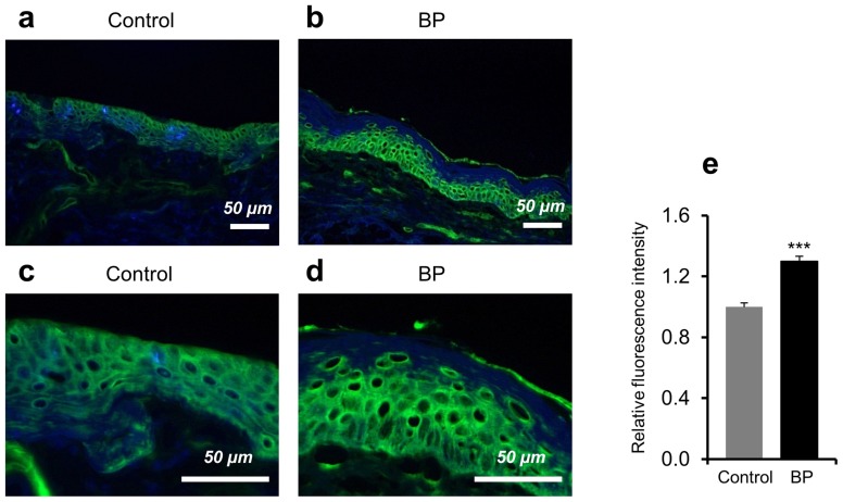
Figure 1. High expression of Hsp90 in the epidermis of bullous pemphigoid patients.
Hsp90 expression was analyzed in perilesional skin biopsies of bullous pemphigoid (BP) patients and in skin sections of healthy controls by fluorescent immunohistochemistry. (a, b) Representative immunofluorescent and DAPI-counterstained (in blue) skin samples showed a stronger intracellular Hsp90 expression (in green) mainly localized to the epidermal layer of the skin of a BP patient compared to control skin. Magnification: x200. (c, d) Higher magnification (x600) of the respective skin samples. (e) Relative mean epidermal Hsp90 fluorescence intensity by densitometry measurements. Values are mean ± SEM of 6 BP patients and 6 controls. ***P<0.001.

