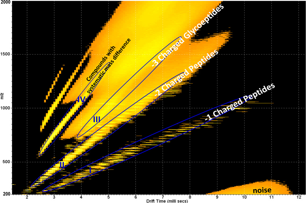
Fig. 9.
2-D IMMS plot of trypsin digested human antithrombin III in negative mode. Trend line I: 2-D region for separation of −1 charged peptides; Trend line II: 2-D region for separation of −2 charged peptides; Trend line III: 2-D region for separation of −3 charged glycopeptides; Trend line IV: Unidentified compounds with systematic mass differences.

