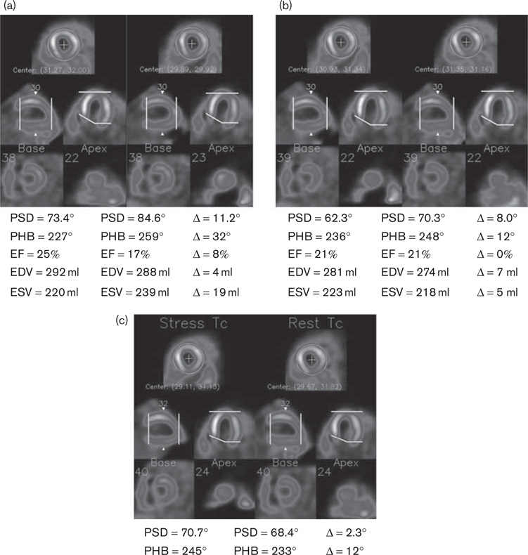
Fig. 2.
A patient example obtained by blinded processing (a), manual side-by-side processing (b), and automatic alignment (c). When processed blinded, the two serial images had inconsistent oblique reorientation [shown on the horizontal long-axis images in (a)] and apical slice selection [shown on the vertical long-axis images in (a)]. Manual side-by-side processing and automatic alignment better aligned the two images and improved the repeatability of the measured parameters. EDV, end-diastolic volume; EF, ejection fraction; ESV, end-systolic volume; PHB, phase histogram bandwidth; PSD, phase standard deviation.

