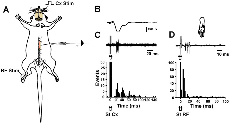
Figure 6. Dorsal horn neurons responding to sensorimotor cortex stimulation also receives input from larger cutaneous afferent fibers.
A, experimental arrangement. B, averaged cortical EFP recorded at a depth of 300 µm (top trace). C, extracellular recording of a single neuron in the same experiment (bottom trace) and peri-stimulus time histogram computed from the action potential responses evoked by cortical stimulation of 10 recorded neurons. D, the trace shows superimposed action potential responses evoked by electric stimulation of the receptive field (RF) (represented in the paw drawing) of the same neuron recorded in C, and peri-stimulus time histogram computed from the action potential responses evoked by the RF stimulation of the same recorded neurons that respond to cortical stimulation. The arrows indicate the cortical (Cx) and RF stimulation time.

