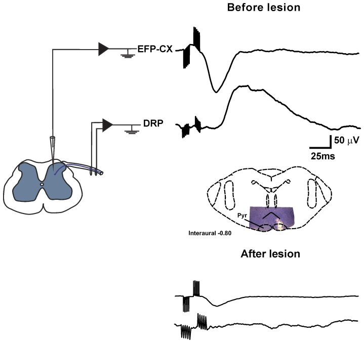
Figure 7. Cortical EFPs and DRPs are suppressed after pyramidal tract lesions.
Averaged EFPs (top traces) and DRPs (bottom traces), evoked by contralateral sensorimotor cortex stimulation, recorded in the L4 spinal cord segment before and after electrolytic lesion of the ipsilateral pyramidal tract at the medullary level. The schematic drawing shows the lesion produced in the pyramidal tract. Notice that both DRPs and EFPs are suppressed after pyramidal tract lesion.

