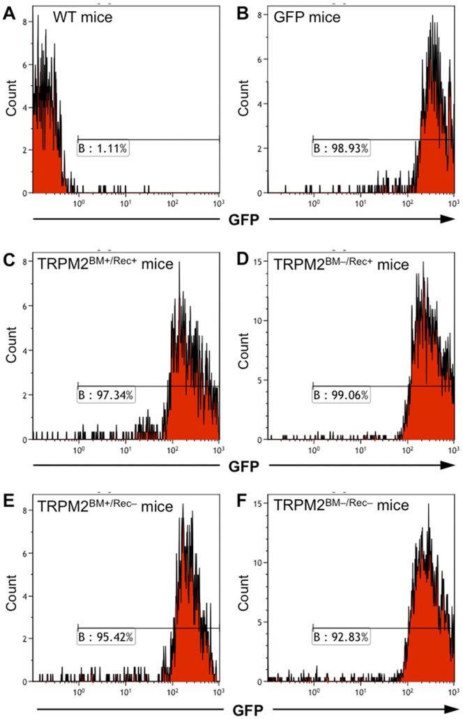
Figure 1. Flow cytometry analysis of BM-derived cells in WT/TRPM2-KO BM chimeric mice.
Representative histograms of GFP+ cells in WT mice (A; negative control), GFP-transgenic mice (B; positive control), TRPM2BM+/Rec+ mice (C), TRPM2BM–/Rec+ mice (D), TRPM2BM+/Rec– mice (E), and TRPM2BM-/Rec– mice (F). In all examined chimeric mice, more than 90% of the BM-derived cells were GFP+.

