
Figure 7. In vivo GLI1 expression after intratumoral administration of NVP-LDE225.
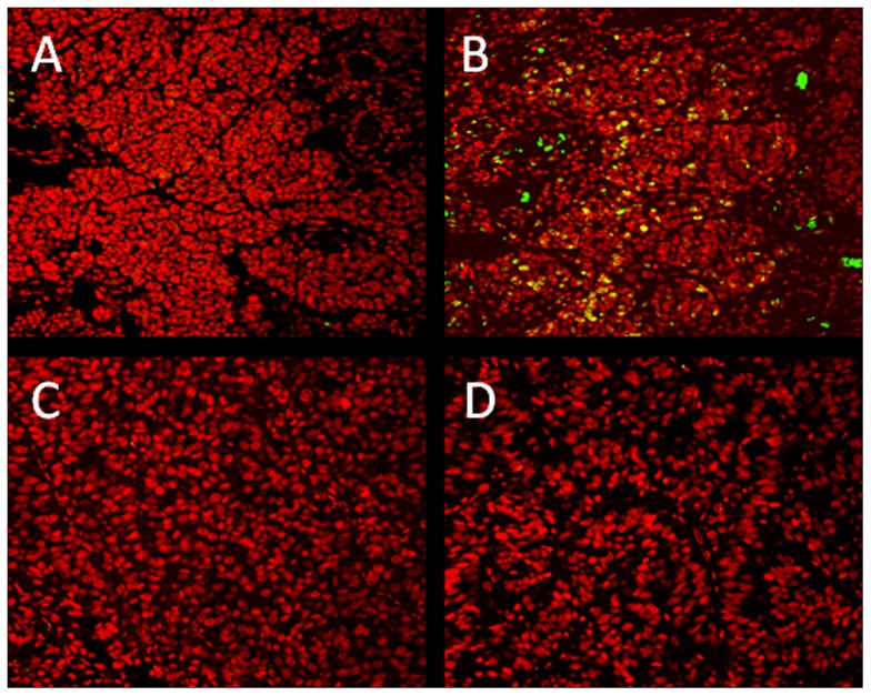
LOX OMVI human melanoma cells were injected s.c into both flanks as mentioned above. Tumors were treated intratumorally on daily basis with vehicle (A & B) or NVP-LDE225 (C & D). Immunofluorescent microscopy of GLI1was performed on isolated tumor tissues. GLI1 staining was performed by overnight incubation of sections at 4°C with rabbit anti-human polyclonal Ab (B & D, NBP1-78259, Novus Biologicals, Littleton, CO) or isotype control (A & C) followed by an 1 hr-incubation with Alexa Fluor® 488 Donkey IgG, anti-rabbit (A21206, Invitrogen, Carlsbad, CA) at RT (green). Counterstaining of nuclei was performed with propidium iodide (red). Pictures were taken on a confocal laser-scanning microscope system (LSM 410; Zeiss). Yellow color corresponds to double positive (anti-GLI1 and propidium iodide) nuclear staining.