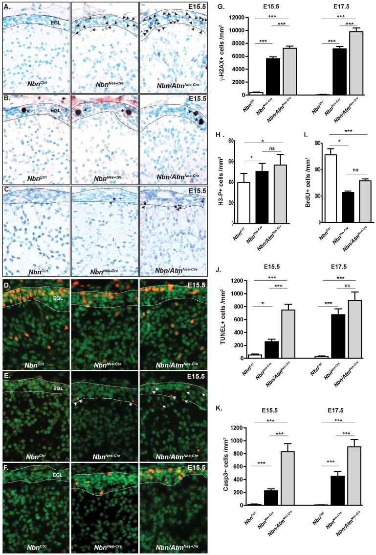
Figure 3. Nbn and Atm inactivation leads to accumulation of DSBs, proliferation defects and apoptosis in the EGL of the cerebellum.
(A) Loss of Nbn and Atm leads to increase of DSBs at E15.5 as indicated by γ-H2AX foci (NbnCtrl, n = 3; NbnNes-Cre, n = 5 and Nbn/AtmNes-Cre, n = 4) (Magnification ×400). (B) Accumulation of neural progenitors in mitosis in EGL primordium as indicated by H3 phosphorylation (Magnification ×400). (C) Cleaved Caspase-3 staining demonstrates increased apoptosis in NbnNes-Cre and Nbn/AtmNes-Cre EGL. (Magnification ×400) (D) BrdU immunohistochemistry shows the reduction in cell proliferation of the neural progenitors in the NbnNes-Cre and Nbn/AtmNes-Cre EGL (Magnification ×400). (E) TUNEL staining demonstrates increased apoptosis in NbnNes-Cre and Nbn/AtmNes-Cre EGL (Magnification ×400). (F) p53 is stabilized in NbnNes-Cre and Nbn/AtmNes-Cre EGL (Magnification ×400). (G) Quantification of cells with γ-H2AX foci in the EGL primordium at E15.5 (NbnCtrl, n = 3; NbnNes-Cre, n = 5 and Nbn/AtmNes-Cre, n = 4) and E17.5 (NbnCtrl, n = 3; NbnNes-Cre, n = 3 and Nbn/AtmNes-Cre, n = 3). (H) Quantification of phospho-histone 3 positive cells (H3-P+) in the EGL primordium at E15.5 (NbnCtrl, n = 4; NbnNes-Cr e, n = 5 and Nbn/AtmNes-Cre, n = 5). (I) Quantification of BrdU positive cells in the EGL primordium At E15.5 (NbnCtrl, n = 5; NbnNes-Cre, n = 3 and Nbn/AtmNes-Cre, n = 4). (J) Quantification of TUNEL positive cells in the EGL primordium at E15.5 (NbnCtrl, n = 4; NbnNes-Cre, n = 3 and Nbn/AtmNes-Cre, n = 4) and E17.5 ((NbnCtrl, n = 3; NbnNes-Cre, n = 3 and Nbn/AtmNes-Cre, n = 4). (K) Quantification of Cleaved Caspase-3 positive cells in the EGL primordium at E15.5 (NbnCtrl, n = 3; NbnNes-Cre, n = 5 and Nbn/AtmNes-Cre, n = 4) and E17.5 (NbnCtrl, n = 3; NbnNes-Cre, n = 3 and Nbn/AtmNes-Cre, n = 4) Error bars indicate SEM. (* p<0,05, ** p<0,01, *** p<0,001, ns: non-significant).

