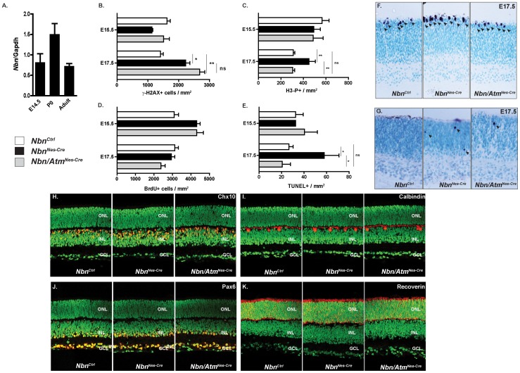
Figure 6. Nbn-deficiency leads to Atm-dependent apoptotic cell death in developing retina, but cell fate specification and differentiation are normal in the Nbn/Atm-deficient retina.
(A) Relative expression of Nbn mRNA at different stages of mouse retinal development was analyzed by real time RT-PCR using SYBR green. No significant difference in the levels of Nbn mRNA expression was detected within the stages analyzed. Gene expression was normalized to Gapdh mRNA (Gapdh). (B) Quantification of γ-H2AX positive cells within retina at E15.5 (NbnCtrl, n = 3; NbnNes-Cre, n = 4; and Nbn/AtmNes-Cre, n = 3) and E17.5 (NbnCtrl, n = 2; NbnNes-Cre, n = 3; and Nbn/AtmNes-Cre, n = 3). (C) Quantification of H3-P positive cells per mm2 of retinal tissue at E15. 5 (NbnCtrl, n = 8; NbnNes-Cre, n = 6 and Nbn/AtmNes-Cre, n = 5) and at E17.5 (NbnCtrl, n = 13; NbnNes-Cre, n = 3 and Nbn/AtmNes-Cre, n = 6). (D) Quantification of BrdU positive cells within retina at E15.5 (NbnCtrl, n = 10; NbnNes-Cre, n = 6; and Nbn/AtmNes-Cre, n = 12) and E17.5 (NbnCtrl, n = 8; NbnNes-Cre, n = 3; and Nbn/AtmNes-Cre, n = 2). Error bars indicate SEM (* p<0,05, ** p<0,01, ns: non-significant). (E) Quantification of apoptotic cells indicated by TUNEL positive cells per mm2 of retinal tissue at E15.5 (NbnCtrl, n = 5; NbnNes-Cre, n = 1 and Nbn/AtmNes-Cre, n = 4) and E17.5 (NbnCtrl, n = 15; NbnNes-Cre, n = 3 and Nbn/AtmNes-Cre, n = 5) (F) Phospho-histone H3 (H3-P) immunohistochemistry of NbnCtrl, NbnNes-Cre and Nbn/Atm Nes-Cre retina at E17.5 (Magnification ×400). (G) TUNEL staining of NbnCtrl, NbnNes-Cre and Nbn/Atm Nes-Cre retina at E17.5 (Magnification ×400). (H–K) Retinal cryosections of P9 retinas from NbnCtrl, NbnNes-Cre and Nbn/AtmNes-Cre were stained with cell type–specific antibodies as shown, in red, for Chx10 (bipolar cells) (H), for calbindin (horizontal cells) (I), for Pax6 (amacrine cells) (J) and for recoverin (photoreceptors and a subset of bipolar cells) (K). Nuclei are shown in green (sytox green counterstaining). Immunohistochemistry patterns of all cell types analyzed are indistinguishable between control, Nbn-deficient and Nbn/Atm-double-deficient retinas (Magnification ×400). Abbreviations: ONL, outer nuclear layer; INL, inner nuclear layer; GCL, ganglion cell layer. Error bars indicate SEM. (* p<0,05, ** p<0,01, ns: non-significant).

