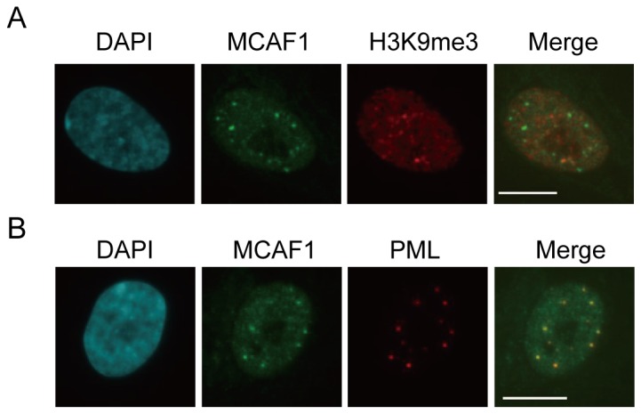
Figure 1. MCAF1 localizes to PML bodies in IMR90 fibroblasts.

(A) Immunofluorescence analysis of endogenous MCAF1 and H3K9me3 in IMR90 cells. DNA was stained with DAPI. Scale bar: 10 µM. (B) Immunofluorescence of endogenous MCAF1 and PML in IMR90 cells. Scale bar: 10 µM.
