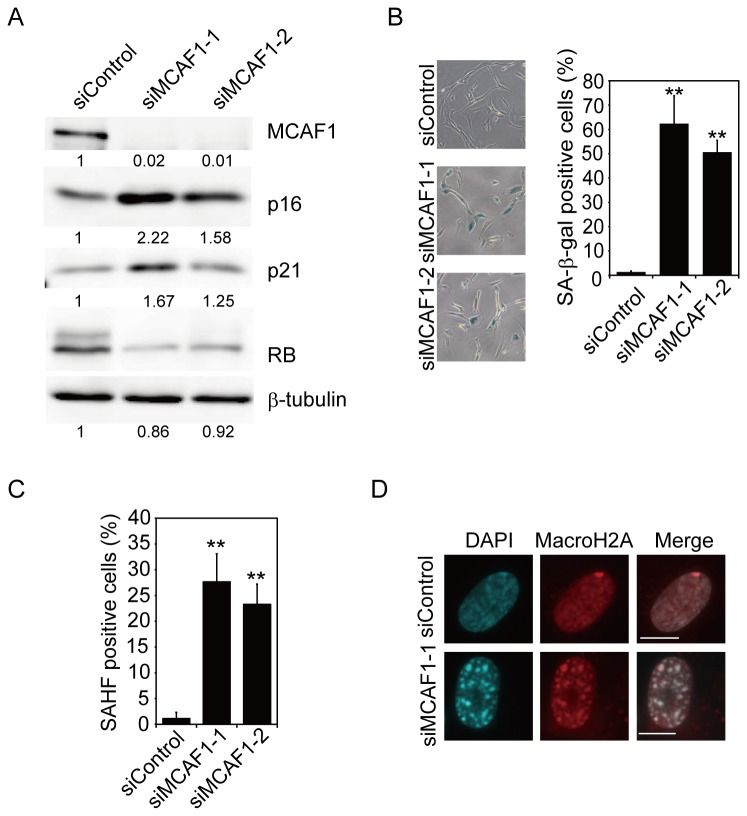
Figure 3. Knockdown of MCAF1 induces premature senescence.
(A) Western blot analysis of the cdk inhibitors p16 and p21 and RB proteins in control and MCAF1 knockdown IMR90 cells. The images were quantitatively assessed by densitometry. (B, C) IMR90 cells were treated with the indicated siRNAs, and analyzed for SA-β-gal activity (B) or the formation of SAHF (C) at 8 days after siRNA treatment. **P<0.01. (D) Immunofluorescence analysis of MCAF1 and MacroH2A in control and SAHF-positive MCAF1 knockdown cells. DNA was stained with DAPI. Scale bar: 10 µM.

