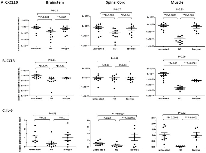
Figure 7. Administration of N3 reduced pro-inflammatory chemokines expression in CNS and muscle tissues of 5746-preinfected hSCARB2-transgenic mice.
RNAs were extracted from the brain, spinal cord, and muscle of 5746-infected 7-day old hSCARB2-transgenic mice treated with N3 or isotype antibody, and antibody-untreated mice as described in Fig. 6 and were subjected to quantitative RT-PCR specific to (A) CXCL10, (B) CCL3, and (C) IL-6. The number of PCR cycles (Ct) required for fluorescent detection of target genes was calculated and presented as the relative expression after normalization with the internal control of β-actin expression from the same tissue. Each normalized 2Ct value was the ratio to the value from the mean of 2Ct obtained from the antibody-untreated tissues. A schematic representation of the target gene expression and the statistical average from 10 mice per group is shown. Unpaired student t test with Welch correction was used for statistical analysis. (*p< = 0.05, **p< = 0.01).

