
Figure 6. GNMT is localized to nuclei.
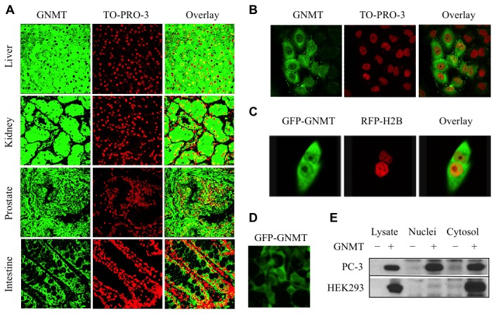
A. Detection of GNMT in a panel of normal human tissues by immunohistochemical staining and confocal microscopy (left panel, GNMT (green) detected by immunostaining with anti-GNMT antibody and secondary antibody conjugated with Alexa Fluor 488 dye; middle panel, nuclei (red) stained with To-Pro-3 dye; right panel, overlay with yellow indicating co-localization). B. Nuclear localization of GNMT in fixed PC3 cells assessed by confocal microscopy: left panel, GNMT (green) detected by immunostaining with anti-GNMT antibody and secondary antibody conjugated with Alexa Fluor 488 dye; middle panel, nuclei (red) stained with To-Pro-3 dye; right panel, overlay with yellow indicating co-localization. C. Accumulation of GNMT in nuclei monitored by confocal microscopy in live cells: left panel, GFP-GNMT (green); middle panel, RFP-H2B (red); right panel, overlay with yellow indicating co-localization. D. Exclusion of GFP-GNMT from nuclei in HEK293 cells detected by fluorescence microscopy. E. Detection of GNMT in nuclear and cytosolic fractions of PC-3 and HEK239 cells 96 h post-transfection. Subcellular fractions were obtained by differential centrifugation. Control (-) or GNMT transfected (+) cell fractions are shown.