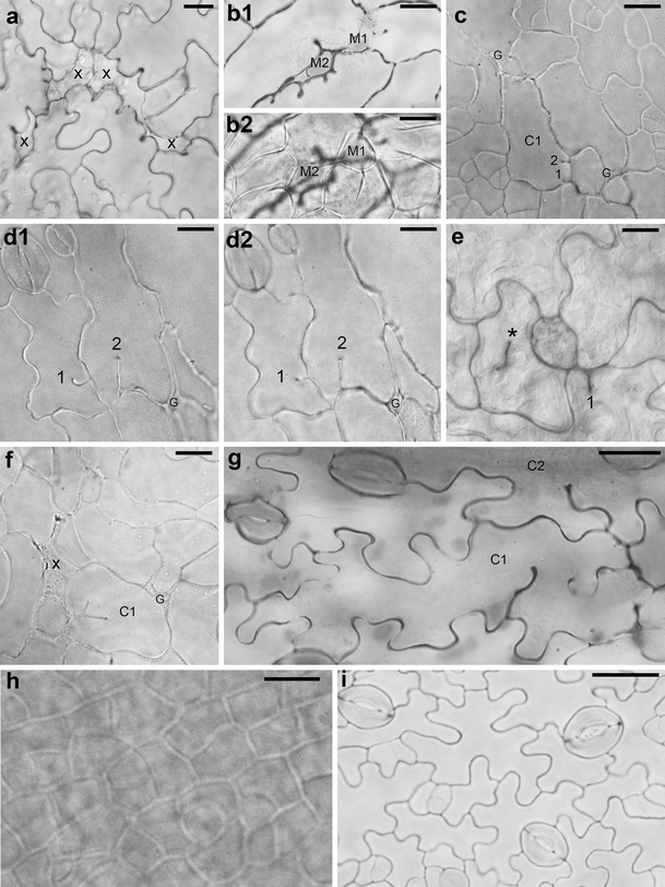
Fig. 1.

a–i Micrographs showing optical sections of abaxial (a, c–i) or adaxial (b) epidermis of cleared whole mount preparations of P STM :CDKA;1.N146 (a–g) and Col-0 (h–i) leaves. a Sites of cell loss (x) surrounded by cells with anticlinal wall invaginations (line B3–4; leaf with lamina c. 1 mm long). b 1, 2 Two optical sections of the leaf fragment. b 1 Through epidermis, cell loss sites that are partly filled by rounded cells (M1 and M2) are shown; b 2 through underlying palisade mesophyll, the M1 and M2 can be recognized as mesophyll cells (B3–5 leaf; c. 4 mm). c Epidermis fragment with not yet well-developed wall waviness showing gaps (G) that are most likely not related to cell loss and a cell (C1) with two short ingrowth-like invaginations (1 and 2) not adjacent to a gap (B3–5, c. 2 mm). d 1, 2 Two optical sections of the epidermis region with two invaginations (1 and 2) not adjacent to a gap (G). The comparison of the sections shows that in d 1, closer to the leaf surface, the invagination 1 is longer than that in d 2 (deeper section) unlike the invagination 2 (B3–5, c. 3.5 mm). e An epidermis fragment showing a defected cytokinesis (asterisk labels the anticlinal wall fragment not attached to anticlinal wall of the mother cell) and an invagination 1 in the vicinity of a putative gap (B3–4, c. 2 mm). f Young epidermis fragment showing a cell (C1) with a branched invagination, a putative cell loss site (x), and a gap (G) (B3–5, c. 2 mm). g Epidermis fragment with well-developed wall waviness showing a cell (C1) with two wavy ingrowth-like invaginations that are not in the vicinity of a gap or cell loss site, and a fragment of a neighboring cell (C2) with a single wavy invagination (B3–5, c. 2.5 mm). h Fragment of wild-type epidermis with dividing cells and wall waviness not yet developed (c. 2 mm). i Wild-type epidermis with well-developed wall waviness and active meristemoids (c. 3.5 mm). Scale bars 20 μm (a, h, i) and 10 μm (b–g)
