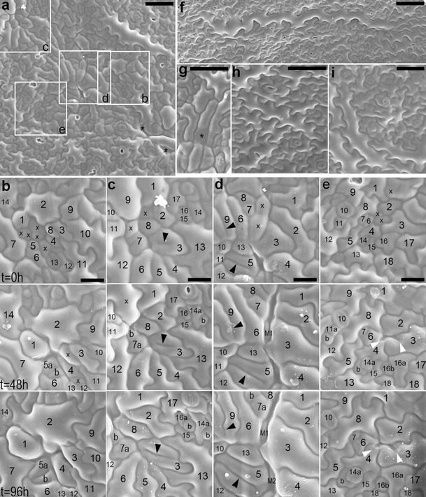
Fig. 2.

a–i SEM micrographs showing abaxial epidermis replicas of proximal portions of B3–5 (a–g) and Col-0 (h, i) leaves. a Epidermis region (the first replica of the sequence) from which fragments outlined by rectangles are shown in b–e (leaf lamina c. 3 mm long). Asterisks mark sites of cell loss. b–e Fragments of epidermis region shown in a. Each column is a sequence of consecutive replicas; time at which they were taken is given on left of each row. In each column, the cells have the same numbers; a cell progeny is labeled with letters; cells are marked with x if they are lost in the next replica. b Five cells are lost after the first replica taking; cell 1 grows so that this space becomes closed. c Cell 3 has an interior fragment of anticlinal wall (arrowhead) not attached to its outer anticlinal walls. Some cells are lost, and their sites are filled by surrounding cells. d Cells 5 and 9 have long ingrowth-like invaginations (arrowheads). In the elongated gaps formed due to cell loss, underlying mesophyll cells (M1 and M2) are appearing. e Anticlinal walls of cells 3 and 4 form invagination (arrowheads) after a neighboring cell loss. A space between cells 2–4 and 6 is closing. f A giant cell surrounded by over 30 much smaller neighbors (leaf c. 6 mm). This cell is more or less parallel to leaf margin, located c. 400 μm from the margin. g Fragment of replica, the first in the sequence, showing a cell (asterisk) which outer periclinal wall has probably collapsed (leaf c. 2.5 mm long). h The second replica taken from wild-type epidermis (leaf c. 3 mm). i The second replica taken from wild-type epidermis of the leaf older than that in h (leaf c. 5 mm). Scale bars 50 μm (a, g–i), 20 μm (b–e), and 100 μm (f)
