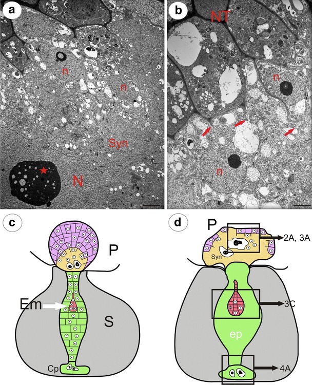
Fig. 2.

Sections through the mature syncytium of Utricularia minor and schematic drawings of the young Utricularia seeds. a Micrograph showing the ultrastructure of the syncytium with two different populations of nuclei; syncytium (Syn), giant endosperm nucleus (N), nucleolus (star), nucleus from nutritive tissue cell (n), bar = 2 μm. b The peripheral part of the syncytium, where the protoplasts of nutritive cells merge with the syncytium; nutritive tissue (NT), arrows—digested nutritive tissue cell walls, nucleus from nutritive tissue cell (n), bar = 1.2 μm. c, d Schematic drawings of two developmental stages of young Utricularia seeds, showing relationships between the endosperm–placenta syncytium, chalazal haustorium, endosperm proper and embryo; placenta (P), seed (S, gray color), nutritive tissue (violet color), endosperm proper (ep, green color), embryo (Em, red color), syncytium (Syn, yellow color), Cp (chalazal endosperm haustorium). d In the endosperm proper, the individual cells were not shown. Note that in the older stage (d), only small amount of the nutritive tissue persists, and the syncytium is fully formed and occupied the place of the nutritive tissue. Framed parts mark corresponding photographs shown in a in this figure and in Figs. 3a, c and 4a
