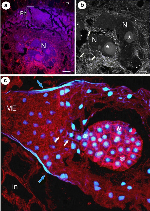
Fig. 3.

Visualisation of microtubules in the syncytium, endosperm and embryo. a Arrangement of the microtubule cytoskeleton in the syncytium of Utricularia intermedia; the peripheral part of the syncytium with nuclei from the nutritive tissue cells (Ph), two giant endosperm nuclei in the central part of the syncytium (N), placenta (P), bar = 36 μm. b The microtubular cage between the lobes of two giant endosperm nuclei syncytium of Utricularia intermedia; giant endosperm nucleus (N), nucleolus (star), additional nucleoli (arrows), bar = 18 μm. c The architecture of the microtubule cytoskeleton in the endosperm proper and embryo of Utricularia minor; micropylar part of endosperm proper (ME), cuticle (arrows), suspensor’s cells (white arrows), integument (In), bar = 10 μm
