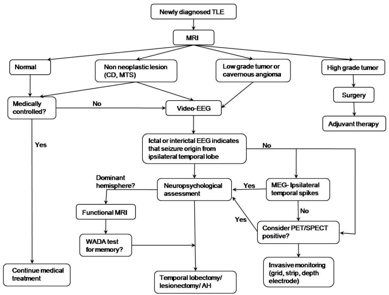
Fig. 2.
Algorithm of epileptic surgery in a patient with temporal lobe epilepsy (TLE). MRI, magnetic resonance imaging; CD, cortical dysplasia; MTS, mesial temporal sclerosis; EEG, electroencephalogram; MEG, magnetoencephalography; PET/SPECT, positron emission tomography/single photon emission computed tomography; AH, amygdalohippocampectomy. Reprinted from Benifla, et al. Neurosurgery 2006;59:1203-13, with permission of Lippincott Williams & Wilkins16).

