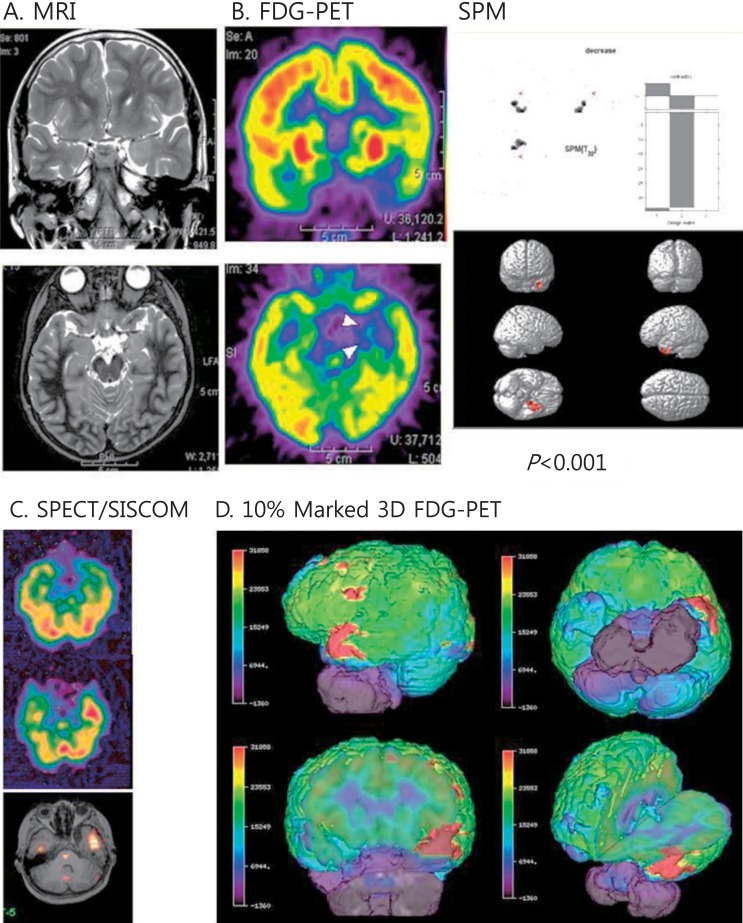
Fig. 3.
Multimodal neuroimages of a patient with left temporal lobe epilepsy due to ganglioglioma. (A) Magnetic resonance imaging (MRI) showed increased signals on the left uncus and hippocampus. (B) Fluorodeoxyglucose-positron emission tomography (FDG-PET) showed decreased glucose metabolism in the same areas. Statistical parametric mapping (SPM) analysis also indicated decreased glucose metabolism in the same areas compared to normal controls (P<0.001). (C) Ictal single photon emission computed tomography (SPECT) and SISCOM (subtraction ictal SPECT coregistered to the MRI) demonstrated a significant increased ictal uptake on the left temporal region. (D) Coregistered surface marked FDG-PET images with a 10% asymmetric threshold to the MRI showed red zones on the left temporal areas in various views.

