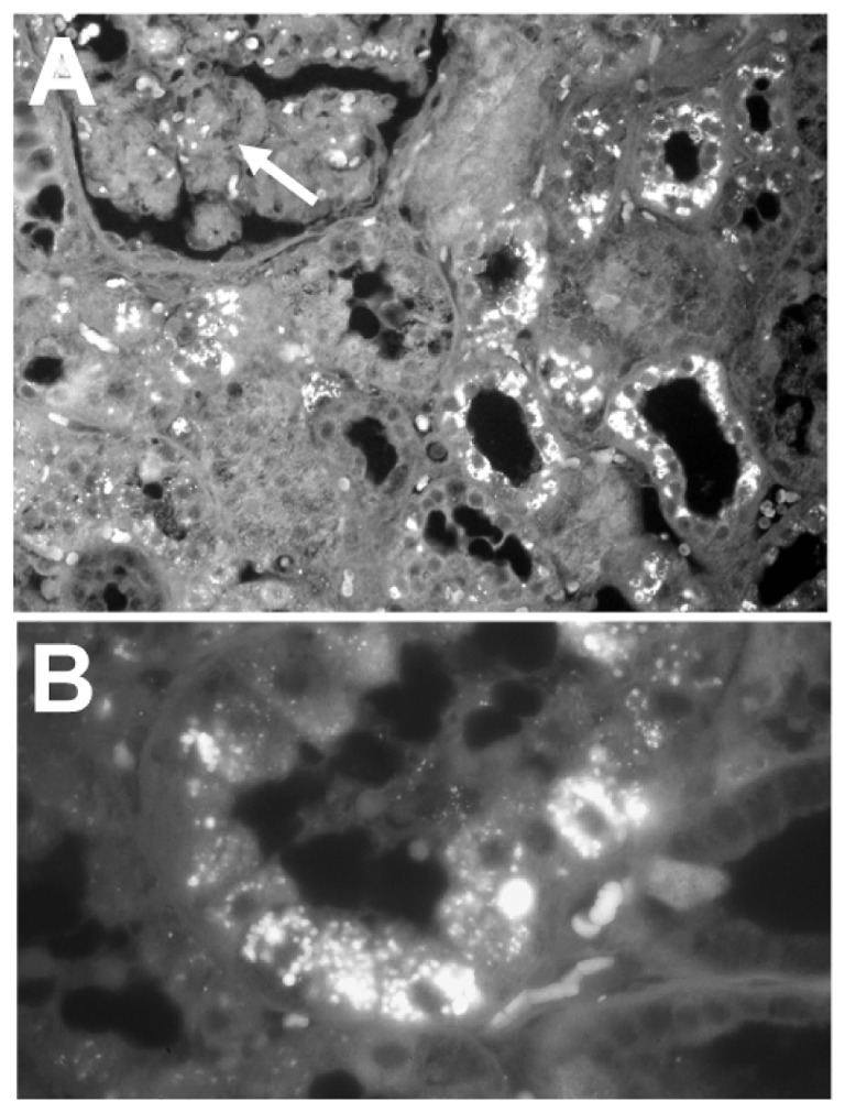
Figure 3.

Immunohistochemical identification of osteopontin in human kidney. A) Part of a glomerulus is identified (arrow). Note the intense positivity in the convoluted tubules on the right. Original magnification: ×50. B) Higher magnification of a section of a convoluted tubule, the OPN granules are identified within the cell cytoplasms. Original magnification: ×200.
