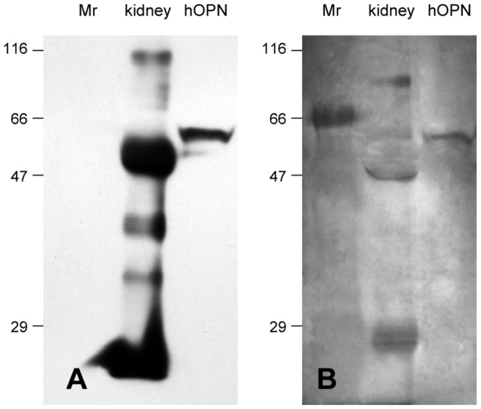
Figure 6.

Human OPN detection in kidney tissue by western blotting (A) and AgNOR staining on the same membrane (B). Kidney cell lysate was immunoprecipitated with anti human OPN peptide (75-90) antibody. Immunoprecipitated complexes and 6.25ng of hOPN control were subjected to 12% SDS-PAGE and western blot analysis was performed with anti-hOPN. The blot reveals an intense 56kDa OPN protein in kidney tissue and a 60kDa purified hOPN control. After immunoblotting, membrane stained by AgNOR exhibited a similar pattern with a 56kDa OPN band in human kidney extract and a 60kDa purified hOPN protein. Protein standards (Mr) indicate the position of the molecular weight markers.
