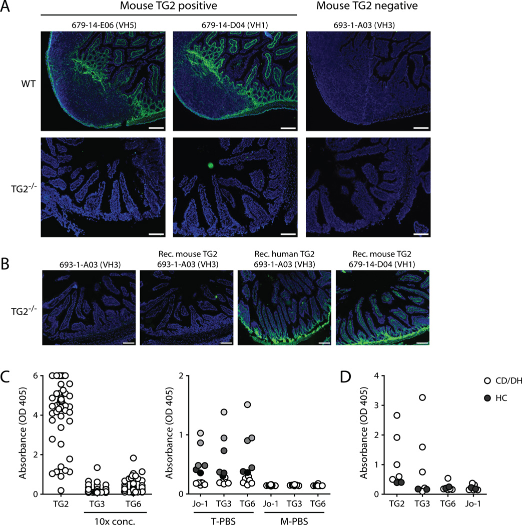
FIGURE 2.
Specificity of TG2-reactive mAbs. (A and B) Representative micrographs showing immunofluorescence staining of mouse intestinal tissue cryosections with TG2-reactive mAbs. Human IgG is shown in green and cell nuclei are shown in blue (Hoechst staining). Scale bar, 100 µm. (A) Staining of wild-type (WT) or TG2 knockout (TG2−/−) (21) mouse tissue with three different mAbs. The wild-type tissue contains a Peyer’s patch. Two of the mAbs reacted with mouse TG2 in ELISA, whereas the third one did not. (B) Reactivity of TG2-specific mAbs with TG2−/− tissue could be achieved by incubating the cryosections with recombinant mouse or human TG2. After washing, added TG2 remains bound to the ECM. (C) The left panel shows the reactivity of 57 mAbs with TG2, TG3 and TG6 in ELISA. Each circle represents a single mAb tested in one concentration. The mAb concentrations used with TG3 and TG6 were 10 times higher than the concentrations used with TG2 as antigen. The right panel shows the reactivity of 12 selected mAbs with the autoantigens Jo-1, TG3 and TG6 in either PBS with 0.1% tween (T-PBS) or PBS with 2% dry milk (M-PBS). The data points are color-coded so that the reactivity of individual mAbs can be followed with different antigens. (D) Reactivity of IgA antibodies in serum of patients with celiac disease and dermatitis herpetiformis (CD/DH) (n=5) or healthy controls (HC) (n=2) with TG2, TG3, TG6 and Jo-1 determined by ELISA. Both treated and untreated patient sera were used. The sera were used in a dilution of 1:20 and incubated with the antigens in the presence of 2% dry milk.

