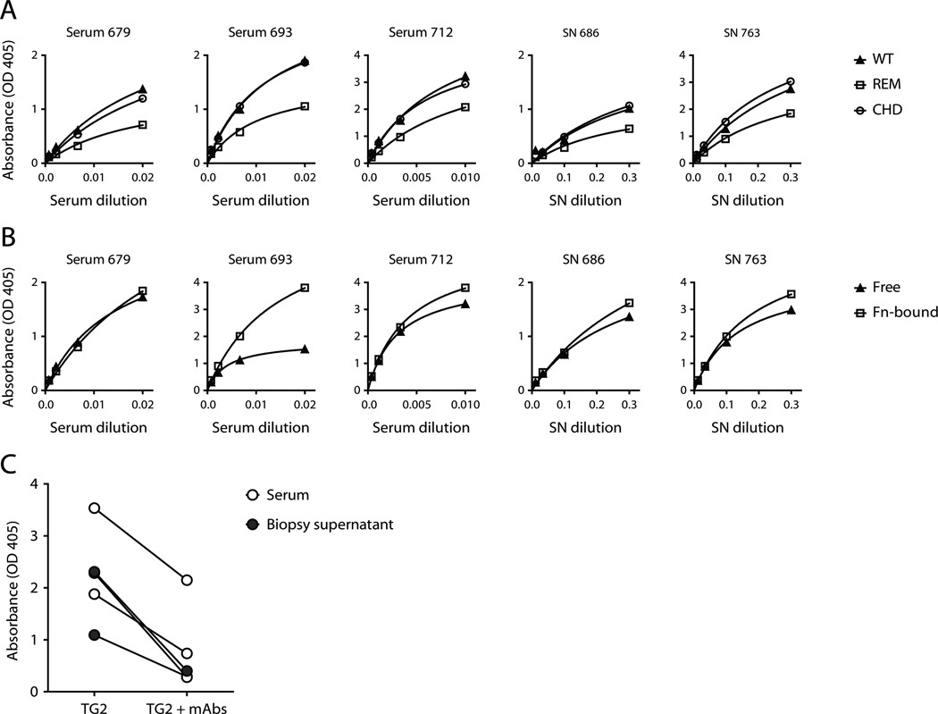
FIGURE 5.
Reactivity of polyclonal IgA from celiac disease patients. (A and B) Saturation binding curves showing the reactivity of polyclonal IgA antibodies with TG2 in ELISA. Celiac disease patient serum and supernatants (SN) from cultured small intestinal biopsies were used as sources of polyclonal IgA. (A) Comparison of reactivity to wild-type (WT) and mutant TG2. The tested mutants were the R19S E153S M659S (REM) triple mutant and the C277A H335A D358A (CHD) catalytic triad mutant. (B) Comparison of reactivity to directly coated TG2 (Free) and TG2 captured on the 45 kDa gelatin-binding fragment of human fibronectin (Fn-bound). (C) Detection of TG2-bound IgA in ELISA after incubation of coated TG2 with a single concentration of serum (n=3) or biopsy supernatant (n=2) in the presence or absence of three pooled IgG mAbs: 679-14-E06, 693-1-A03 and 763-4-A06, representing epitope 1, 2 and 3, respectively.

