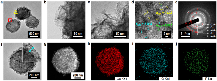
Figure 3. TEM characterization.
(a–c), TEM images of the bacteria-supported, hierarchical, porous-Co3O4 superstructures. Low- (a) and high-magnification micrographs (b). The images shown in (b) and (c) are open triangle (yellow line) and square (red line) regions, respectively, located in (a). The highly porous, hierarchical structures can be identified by the assembly of Co3O4 nanocrystals grown on the bacterial template. (d), HR-TEM image of ~ 2–10-nm nanocrystalline Co3O4 grains in the obtained hierarchical, porous-Co3O4/bacteria product. (e), The SAED pattern for the polycrystalline Co3O4 in the hierarchical, porous-Co3O4/bacteria sample. (f), An individual TEM image of the bacteria-supported, hierarchical, porous-Co3O4 sample. The inset shows a 3D-hierarchical Co3O4 layer grown on a bacterial surface (scale bar represents 50 nm). (g–j), The HAADF STEM image (g) and the EDS elemental mapping analysis (h–j) for an individual sample of the bacteria-supported, hierarchical, porous-Co3O4 sample shown in (f), showing the uniform distribution of Co3O4 nanostructures and the biosorption of cobalt ions onto a phosphate functional group associated with the negative charge of glycopolymers (see figure 1b, TA and LTA) in a bacterial cell wall.

