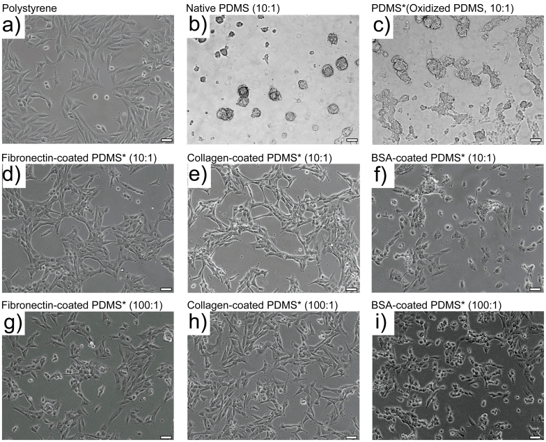
Figure 3. Morphologies of SUM159 colonies on polystyrene and PDMS substrates.
Tumorigenic breast cancer cell line propagated over 2 days on (a) commercial polystyrene petri dish; (b) native (10:1) PDMS; (c–f) plasma-oxidized (10:1) PDMS that were either unmodified (c), fibronectin coated (d), collagen coated (e), or BSA coated (f); (g–i) plasma-oxidized (100:1) PDMS that were either fibronectin coated (g), collagen coated (h), or BSA coated (i). Scale bar: 20 μm. PDMS substrates that were plasma-oxidized are marked with “*” in the images. All ratios are of base to curing agent.

