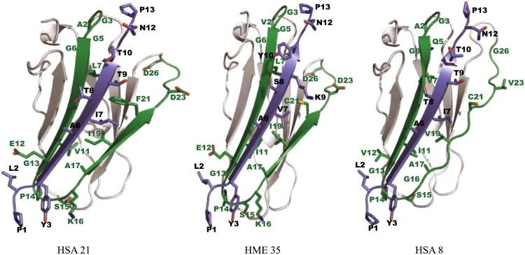
Fig. 7.
Structural modeling of filamin and integrin. Filamin domains are colored grey in non-binding regions and green in the binding region (known as the CD β-strands). Cytoplasmic tails of β-integrins are shown in purple. Positions in the binding filamin pattern (LOGO) with high information content are shown as sticks, with residue names and pattern positions in green. Positions in the integrin pattern with high information content are shown as sticks, with residue names and pattern positions in black.

