Figure 4.
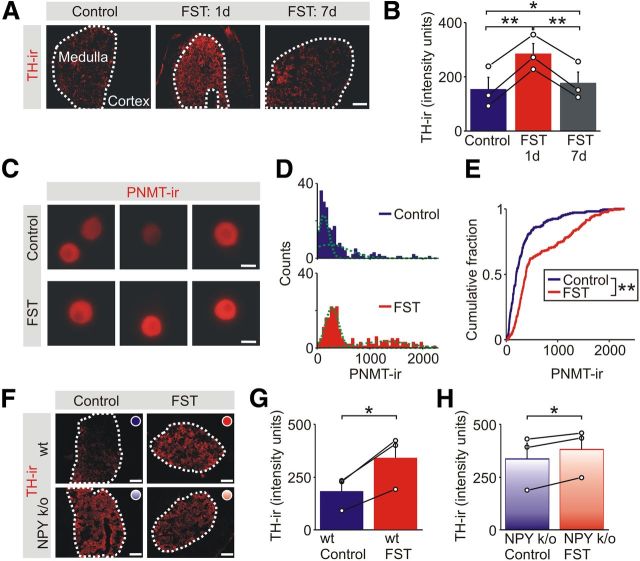
Acute stress leads to a lasting increase in the adrenal expression of enzymes in the catecholamine synthetic pathway. A, TH-IR in adrenal sections from a control animal and 1 d and 1 week after exposure to the FST. Scale bar, 100 μm. B, Group data (mean ± SEM, n = 3) shows that the stress-induced increase in TH-IR in the adrenal medulla that was evident 1 d after the FST had declined toward basal levels by 1 week. C, Examples of PNMT-IR in chromaffin cells from a control animal and 1 d after exposure to the FST. Scale bars, 10 μm. D, Frequency histograms of the levels of PNMT-IR in chromaffin cells from matched control and stressed animals (211 cells for each distribution, n = 3 separate experiments). The histograms were each fitted with two Gaussian distributions (green lines). E, Cumulative frequency distributions showing that the FST led to a significant increase in the levels of PNMT-IR (211 cells for each distribution; Kolmogorov–Smirnov test). F, TH-IR in adrenal sections from control (wild-type or NPY knock-out) mice and 1 d after exposure to the FST. Scale bars, 100 μm. G, In wild-type mice, the FST increased the level of TH-IR compared with matched controls. H, The FST significantly increased the level of TH-IR in the NPY knock-out (k/o) animals, but the increase was smaller than that seen in the wild-type animals. *p < 0.05, **p < 0.01.

