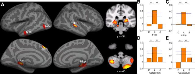
Figure 2.
A, MVPA revealed significant correlations between GMV and form-sound association mapped onto to the inflated gray matter surface as well as in coronal and axial slice. The bar graphs show the prediction accuracy in four example ROIs, including (B) left hippocampus, (C) right hippocampus, (D) left cerebellum, and (E) right cerebellum. Permutation-based 95th percentile (dotted blue line) and 99th percentile (solid blue line) prediction accuracies are also shown (**p < 0.01). Images are reversed left to right to follow radiologic convention. See Table 5 for ROI abbreviations.

