
Figure 12.
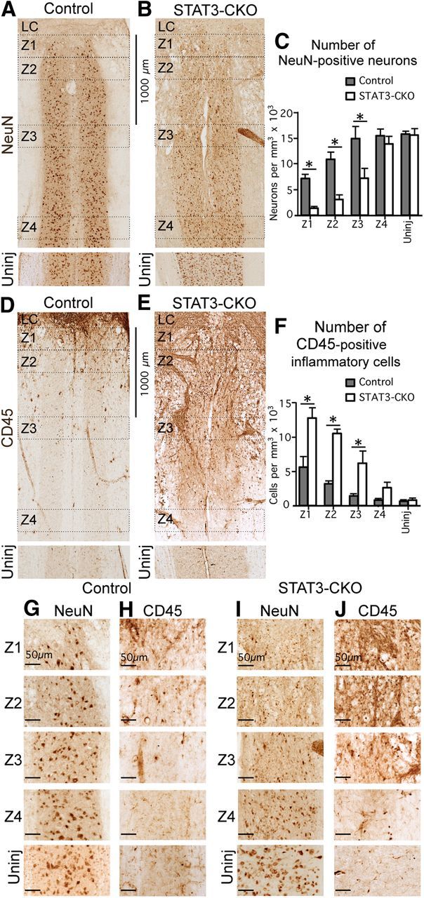
In mice with selective deletion of STAT3 from astroglia, the density of neurons is significantly lower and the density of inflammatory cells is significantly higher in tissue adjacent to the SCI lesion core. A, B, D, E, Bright-field immunostaining showing survey images of NeuN (A, B) and CD45 (D, E) in Z1–Z4 adjacent to the central lesion core and lesion core (LC) at 14 d after SCI in control (A, D) and STAT3-CKO (B, E) mice. C, F, Graphs showing the Neu-N-positive neurons (C), and CD45-positive globoid inflammatory cells (F) in Z1–Z4 after SCI and in uninjured tissue in control and STAT3-CKO mice (n = 4 per group). *p < 0.05 (ANOVA with post hoc pairwise Bonferroni comparison). G–J, Detail images of NeuN-stained neurons (G, I) and CD45-stained inflammatory cells (H, J) taken from survey images in A, B, D, and E. Note the greater loss of neurons and greater number of inflammatory cells in Z1–Z3 in STAT3-CKO compared with control mice. Scale bars: A, B, D, E, 1000 μm; G–J, 50 μm.