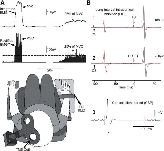
Figure 1.
Experimental setup. A, Raw EMG traces showing (top left traces) with the index finger into abduction by activating the FDI muscle and the visual display presented to all subjects (top right traces) during testing. Subjects were instructed by an oscilloscope to maintain at rest and to perform 25% of MVC with the index finger into abduction. Schematic of the experimental setup showing the posture of both hands and TMS coil during testing (illustration). Note that control subjects completed the test with the right dominant hand and patients with SCI used their less affected hand. B, Raw MEP traces elicited by TMS and TES stimulation recorded from the FDI muscle in a representative subject during all conditions tested. MEPs elicited by the TS (black traces) and CS (red traces) are indicated by arrows during testing of LICI using TMS (B1) and TES (B2). Note that during testing of LICI the CS was given 100 ms before the TS (B1, B2). An example of the CSP (B3) elicited by using TMS during 25% of MVC is presented. The CSP was measured between the stimulus artifact (left dotted line) and the return of background EMG (right dotted line).

