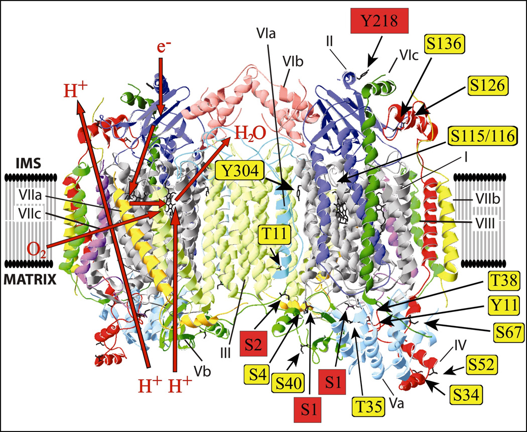
Fig 5.
Identified phosphorylation sites in the crystal structure of bovine heart COX [48]. Crystal structure data of cow heart COX [48] were used and processed with the program Swiss- PDBViewer 4.0.3. Identified phosphorylated amino acids in mammals are represented as sticks. The four newly-identified phosphorylated amino acids are highlighted in red. The red arrows on the left side represent the pathways of hydrogen ions, electrons, dioxygen and water.

