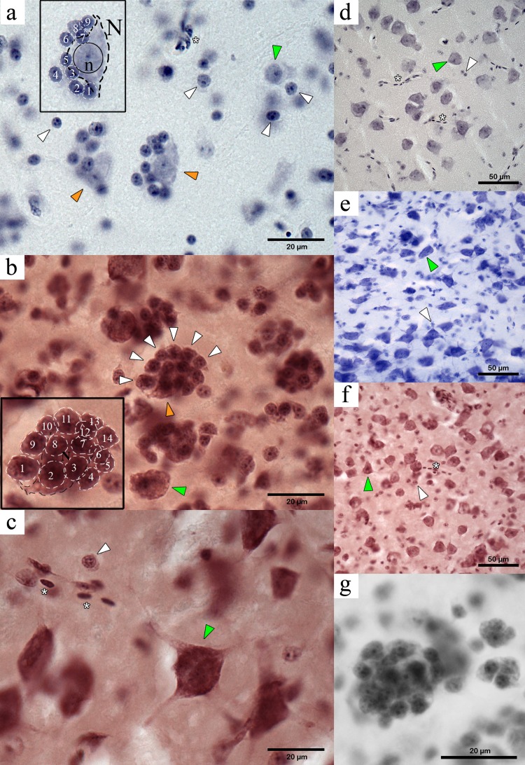
Figure 2. Microphotographs of PGCs in the NC crow telencephalon. Orange arrowheads indicate neurons within a PGC and green arrows indicate unclustered neurons. White arrowheads show perineuronal glia, and the asterisks indicate presence of blood vessels or blood cells.
(A) Medium-sized PGC in the hyperpallium (light haematoxylin stain). Inset shows a large neuron (N) with its dendritic projections (visible contour indicated by dashed black line) showing a round nucleus (solid black circle) with a darkly stained nucleolus (n) in its centre, surrounded by nine perineuronal glia (white dashed lines) (B) Large PGC in the mesopallium (haematoxylin neutral-red stain). Inset showing the visible contour of the central neuron (black dashed line) and of 14 surrounding glia (white dashed line), neurons devoid of a glial cluster is also seen (green arrowhead); (C) Neuron in the hyperpallium apicale devoid of perineuronal cluster (green arrowhead) and isolate glia (white arrowhead). (D) Neurons and glia in the area parahippocampalis (dark haematoxylin stain) with no indication of perineuronal clustering. (E) Neurons (green arrowhead) and oligodendrocytes (white arrowhead) showing no clustered arrangement found in area corticoidea dorsolateralis (cresyl violet stain); (F) Unclustered neurons and glia in the arcopallium (haematoxylin neutral-red stain); (G) Large PGC in the mesopallium (haematoxylin neutral-red stain).

