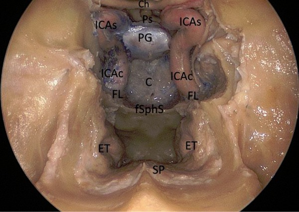
Figure 1:

Endoscopic endonasal anatomic picture showing the suprasellar, the parasellar and the clival area.
Ch: chiasm; Ps: pituitary stalk; PG: pituitary gland; C: clivus; fSphS: floor of the sphenoidal sinus; ET: Eustachian tube; SP: soft palate; FL: foramen lacerum; ICAc: paraclival segment of the internal carotid; ICAs: parasellar segment of the internal carotid artery.
