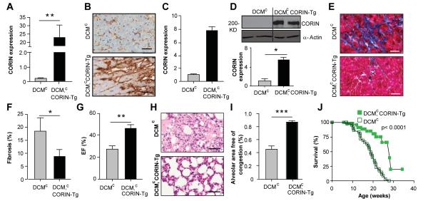
Figure 4. Corin over-expression in DCMc mice reduces fibrosis, HF and increases survival.
(A) Cardiac expression of corin transcripts in DCMc and DCMc, corin-Tg mice assessed by qRT-PCR analysis, relative to wild-type. Transcripts are means of averages of triplicate measures in 7 mice. (B, C) Corin protein expression assessed by immunohistochemical staining. Representative immunoperoxidase-stained heart sections (n= 2 per group) probed with anti-corin antibody (40x magnification, bar = 50 um). Quantification of corin expression by image analyses. (D) Corin protein expression in heart assessed by Western blotting under reducing conditions with anti-corin and anti-actin antibodies. Relative corin levels normalized to actin. (E, F) Cardiac fibrosis in in representative heart ventricles sections (n=4-5 each group) stained with Masson’s trichrome (E, 40 × magnification, bar = 50 um). Quantification (F) of fibrosis by image analyses. (G) Cardiac EF% (n=6-7 per group). (H, I) Alveolar congestion in representative formalin-fixed lung sections (H, 40 × magnification, bar = 50 um) stained by hematoxylin and eosin from female DCMc and DCMc, corin-Tg mice. Bar graph (I) of total alveolar area free of edema and congestion per 20X field. Results are means of averages of 10 randomly selected fields from 6-7 mice of each group. (J) Kaplan-Meyer survival curves of DCMc (n= 56) and DCMc, corin-Tg (n=46) mice. *p≤0.05, **p≤0.01, *** p≤0.001.

