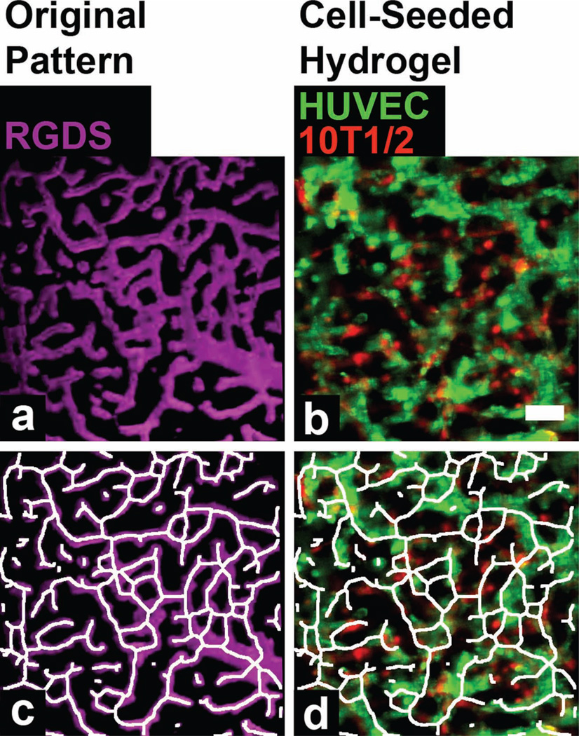
Figure 3.
(a) A degradable PEG-PQ hydrogel with a fluorescent PEG-RGDS pattern (magenta) mimicking the vasculature from the cerebral cortex. (b) HUVECs (green) and 10T1/2s (red) have formed intricate tubule networks that after 24 hours align with the PEG-RGDS pattern of the cerebral cortex vasculature. (c) An artificial skeletonized tracing to highlight the pattern structure. (d) The skeletonized tracing of the pattern has been overlaid with the organized HUVECs (green) and 101/2s (red) to demonstrate excellent alignment of the tubules to the patterned structure. Scale bar = 50 µm.

