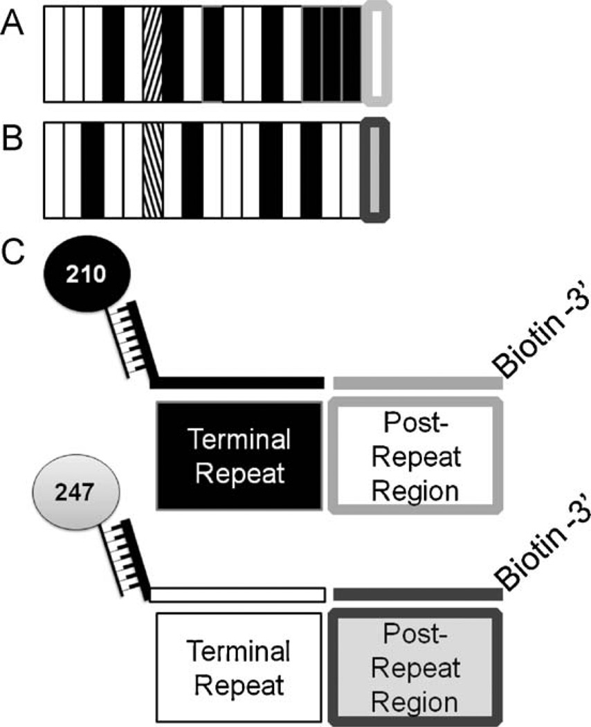
Fig. 2.
Post-PCR ligation detection reaction-fluorescent microsphere assay (LDR-FMA) design. Plasmodium species and pvcsp strain infection were analyzed using post-PCR LDR-FMA. Species diagnosis was performed as previously described in McNamara et al. (2006). P. vivax strain diagnosis based on variants VK210 and VK247 were performed as shown here. (A) VK210 pvcsp repeat region genetic structure. White bars represent ‘A’ repeat motifs, black bars – ‘B’ repeat motifs, and diagonal bars – ‘C’ repeat motif. The conserved post-repeat region is shown as a white box outlined in light grey. These A, B, and C motifs as well as post-repeat sequences are unique to VK210. (B) VK247 pvcsp repeat region genetic structure. White bars represent ‘D’ repeat motifs, black bars – ‘E’ repeat motifs, and diagonal bars – ‘F’ repeat motifs. The light grey box outlined in dark grey represents the conserved post-repeat region. These D, E, and F motifs as well as post-repeat sequences are unique to VK247. (C) Illustrates the LDR primer design capturing the terminal repeat and post-repeat region unique to each P. vivax strain.

