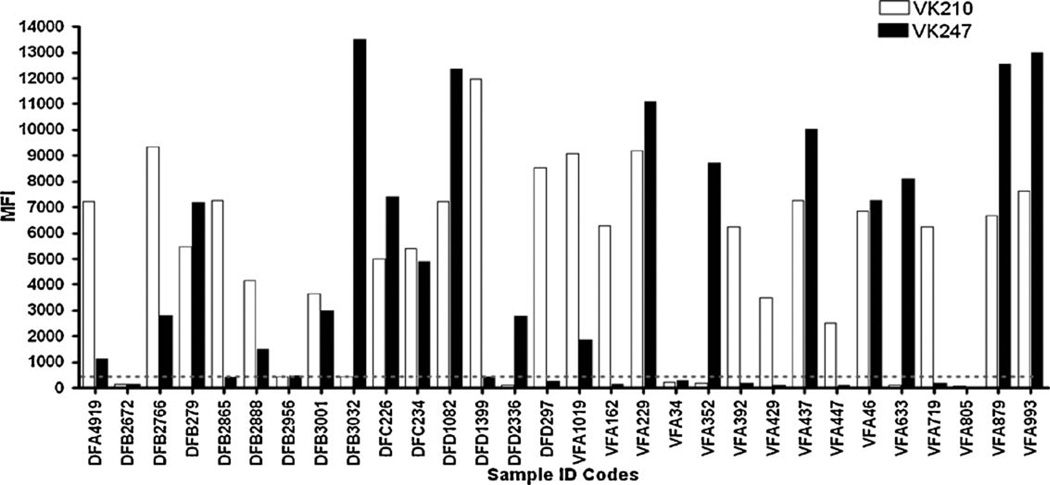
Fig. 4.
Overview of P. vivax VK210 and VK247 strain infections in select Wosera and Mugil study participants. Strain specific fluorescence data shown as median fluorescence intensity (MFI) for VK210 (white) and VK247 (black). Select study participants (n = 20) were from villages in the Wosera (DFA, DFB, DFC, DFD; n = 15) and villages in Mugil (VFA; n = 15) study sites. LDR-FMA MFI values are found on the Y-axis and range from 0 to 14,000. A fluorescence value of approximately 500 (as shown by the grey dotted line) is the threshold for positivity and corrects for background activity. Samples chosen represent the infection status commonly observed in the two populations surveyed. Single infection with VK210 and VK247 are found as well as mixed infection with both variants and lack of P. vivax infection.

