
Figure 1.
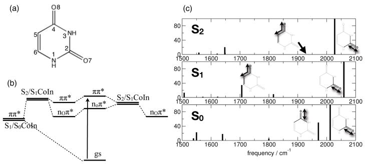
(a) Chemical structure of uracil. (b) Schematic of the femtosecond excited state relaxation mechanisms. (c) Carbonyl stretch vibrations: frequencies and relative intensities of the carbonyl marker bands in the electronic ground state S0 (ν̃C=O7 = 2012 cm−1, ν̃C=O8 = 1971 cm−1), and the excited states S1 (nO8π*; ν̃C=O7 = 2059 cm−1, ν̃C=8 = 1706 cm−1) and S2 (πOπ*; ν̃C=O7 = 2028 cm−1, ν̃C=O8 = 1929 cm−1).