
Figure 1.
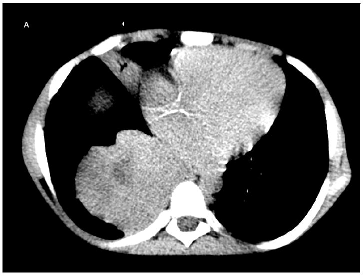
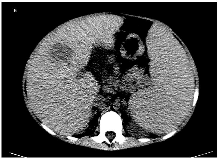
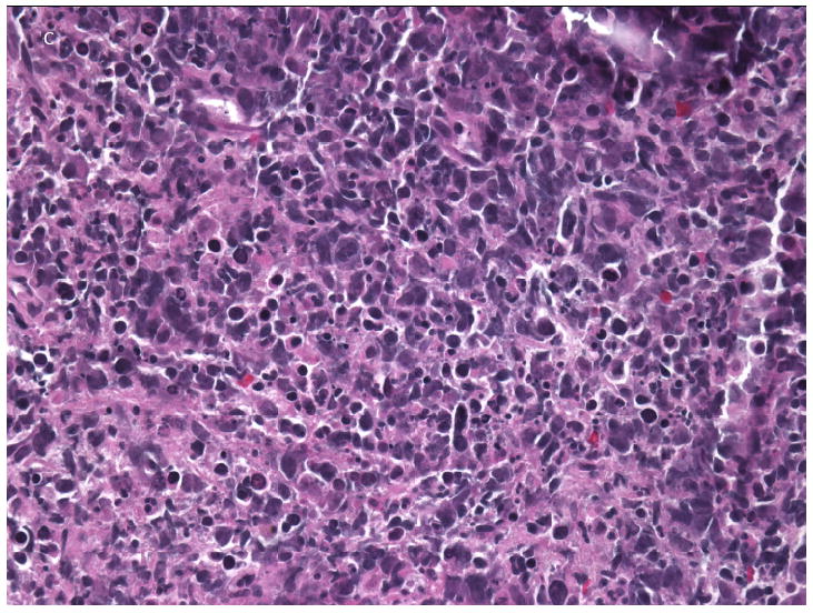
A: Non-contrast chest CT showing extensive consolidation in the right lower lobe with central hypodensity consistent with necrosis. B: Non-contrast abdominal CT showing a large necrotic lesion in the anterior right lobe. C: Liver core biopsy from the necrotic area with pleomorphic lymphoid infiltrate composed of large cells with necrotic debris and frequent apoptotic bodies.