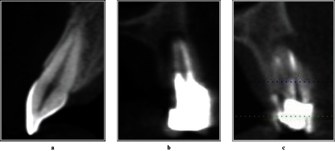
Figure 1.
Illustrative images of the criteria for presence of apical periodontitis (AP) in cone beam CT transversal slices. (a) Healthy tooth with no pathological periapical changes. No hypodense image is observed and the cortical bone is intact. (b) Root-filled tooth with hypodense image of AP between the root limit and periapical bone. (C) Root-filled tooth with hypodense image of AP with buccal cortical destruction

