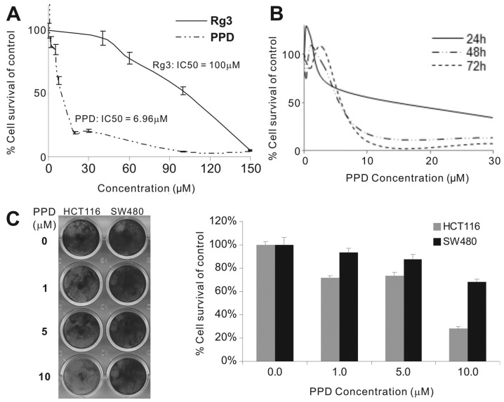
Figure 1.
Effect of protopanaxadiol (PPD) on the proliferation of human cancer cells. (A) MTT assay. HCT116 cells were seeded in 24-well plates and treated with different concentrations of PPD and Rg3 for 48 h. Cells were fixed and subjected to MTT assay. Each treatment condition was carried out in triplicate. (B) Crystal violet assay. HCT116 cells were treated with PPD at the indicated concentrations for 24, 48 and 72 h. Treated cells were subjected to crystal violet staining, which was subsequently dissolved for quantitative readings. Each assay condition was carried out in triplicate. (C) Crystal violet assay in HCT116 and SW480 cell lines. HCT116 and SW480 cells were treated with PPD at the indicated concentrations for 72 h. The gross images (left panel) and quantitative analysis (right panel) of crystal violet staining were obtained. Each assay condition was calculated in triplicate.

