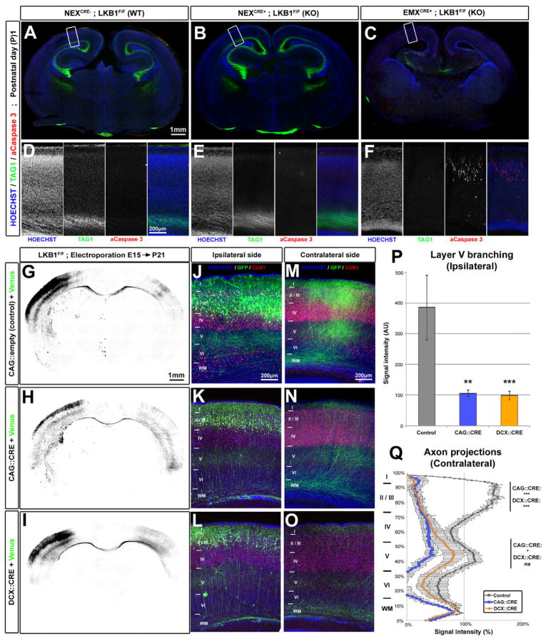
Figure 1. LKB1 deletion after axon initiation does not impair axon maintenance but reduces axon branching in vivo.
(A–C) Coronal section of newborn LKB1F/F mouse brains not expressing Cre recombinase (Wildtype WT, A) or expressing Cre under the NEX (KO) (B) or EMX (KO) (C) promoters. (D–F) Higher-magnification images of the cortex region boxed in A, B and C respectively. (G–I) Low magnification images of coronal brain sections of P21 Lkb1F/F mice electroporated at E15 with plasmids expressing mVenus alone (G) or co-expressing Cre recombinase (under CAG promoter, H or Doublecortin promoter, I) and mVenus. (J–O) Higher magnification of the ipsilateral (J–L) or the contralateral side (M–O) showing reduced axon branching in both Cre-electroporated neurons (K–O) compared to control (J, M). (P–Q) Quantification of normalized Venus fluorescence in layer 5 of the ipsilateral cortex (P, ±SEM) and along the radial axis of the cortical wall in the contralateral cortex (Q, ±SEM). Statistical analysis: Mann-Whitney (P) or two-way ANOVA (Q). See also Fig. S1 and S2.

